Skip to content
Menu
About
Meet Dr. Gallinger
Meet Dr. Trujillo
Meet the Team
Join Our Team
Services
Family Dentistry
Dental Implants
Preventive Dentistry
Restorative Dentistry
Emergency Dentistry
Oral Surgery
Cosmetic Dentistry
Specialty Services
Patients
Frequently Asked Questions
Education
Periodontics
Payment Options
CareCredit
New Patient Specials
Refer a Friend
Blog
Contact Us
Services
Blogs
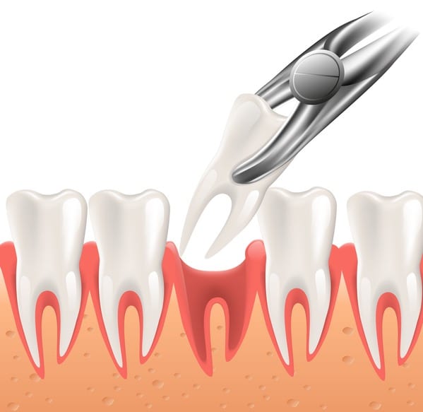
Surgical Instructions
July 09, 2019
Blogs
Oral Hygiene Aids
June 19, 2019
Blogs
Home Care
May 01, 2019
Blogs
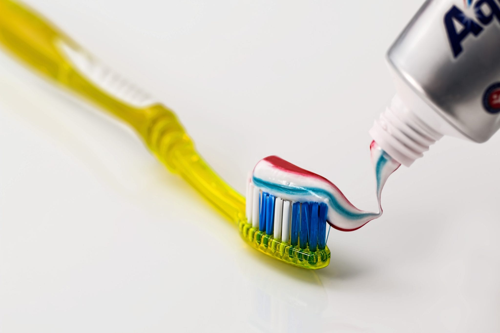
How to Properly Brush & Floss
March 08, 2019
We use cookies to enhance your browsing experience and analyze our traffic. By clicking "Accept", you consent to our use of cookies.
Accept
Privacy policy